


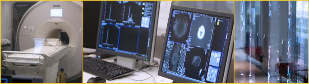
The Center for Image Acquisition (CIA) has a 3 Tesla, Siemens Magnetom Prisma MRI system and a 7 Tesla, Siemens Magnetom Terra MRI system. These scanners are among the most advanced imaging devices approved for use on human subjects, and with them, the CIA and related researchers push the boundaries of possibility in terms of spatial and temporal scans of the brain. Both foundational research and clinical scans are performed at the CIA and the facility is equipped with knowledgeable staff as well as the latest in advanced equipment.
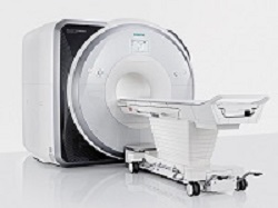
3T Siemens Magnetom Prisma MRI System
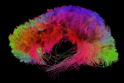
The Prisma 3T is a high performance MRI system based on the Trio system. It allows for unparalleled simultaneous 80mT/m @ 200T/m/s gradients. Its 60 cm bore diameter allows for magnet homogeneity at 40 cm DSV – 0.2 ppm. With 64 – 128 channel receiving, it is the ultimate MRI system for neuroimaging. The Prisma offers standard Diffusion Spectrum Imaging up to 514 diffusion directions, allowing characterization of crossing fibers. It is also capable of higher resolution DTI with a reduced field of view – ZOOMit DTI for isotopic DTI. Higher SNR also allows for reproducible 3-Dimensional, Arterial Spine labeling (ASL) perfusion and improved resting state fMRI data.
The Magnetom Prisma MRI system is FDA-approved, which allows its high-resolution capabilities to be used in the clinical imaging of patients with neurological diseases and other conditions affecting the brain. The key aspect of this system that is unique is its powerful XR Gradients, which have a strength of 80 milliTesla per meter (mT/m) combined with a 200T-per-meter-per-second (T/m/s) slew rate. Furthermore, it employs advanced homogeneity and shimming functions for optimal MRI data acquisition.
3T Technical Specifications
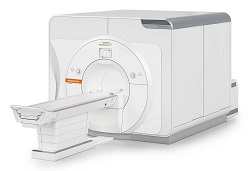
7T Siemens Terra MRI System
The Siemens MAGNETOM Terra is the first 7T MRI scanner for diagnostic imaging and is designed for unprecedented breakthroughs in clinical care. This advanced ultra-high-field (UHF) technology is an actively shielded whole body highly homogeneous superconducting magnet, which was installed at the CIA in March 2017. This unique scanner is designed for both research and clinical applications and can be switched to clinical diagnostic imaging tasks with a Dual Mode feature in less than 7 minutes. A comparison of the two modes is available in the table below.
| RESEARCH MODE | CLINICAL MODE |
|---|---|
| syngo MR E11 software line | syngo MR E11 software line |
| XR gradients 80/200 | XR gradients 80/200 |
| Up to 64 receiver channels | Up to 64 receiver channels |
| MaRS computer | MaRS computer |
| 8 channel parallel transmit | 1 transmit channel |
| 8 x 2 kW RF power | 11 kW RF power |
| Wider range of RF coils | 2 coils available (head and knee) |
The Terra 7T utilizes an actively-shielded, highly homogeneous superconducting magnet housed in an 830 mm horizontal room temperature bore, low-loss helium cryostat, and zero helium boil-off technology. Field shimming is primarily accomplished using superconducting shim coils. Final shimming is performed with a small number of passive shims. This scanner is supplied with a helium level monitor and an emergency quench heater control unit. A two-stage 4.2 K cryo refrigerator cooling system is employed, consisting of two independent cryocooler systems that eliminate the static cryogenic consumption.
 Gradients & RF Technology
The gradient system essentially consists of a Gradient Power Amplifier (GPA) and a Gradient Coil (GC), both water-cooled to sustain a high duty cycle. The Magnetom 7T uses the GPA model similar to that used in other Magnetom MRI systems. The XR gradients are the same as the Prisma, capable of 80mT/m @ 200T/m/s gradients. The gradient coil includes the full set of five second-order electrical shim coils to adjust static magnetic field homogeneity for subject or patient and each measurement volume. The Siemens RF receiver technology uses 64 channels, and the 7T Terra at USC includes a Nova Medical 32-channel head coil. This MRI system also has an 8-channel parallel RF transmit array (pTx). Third order shims provide four out of the seven possible third order shims built into the SC72 gradients, along with second 4-fold SPS cabinet, wiring for third order shim, electric infrastructure expansion, and an SW interface to drive additional third order shims.
The 8Tx-32Rx head coil is the very latest of Nova Medical's high-performance products for the Siemens Whole-body 7T MRI scanner. It features a new highly efficient 8Tx transmit shell. By enabling B1 shimming or full parallel transmission (pTx) operation, the new 8Tx array allows greatly improved B1 transmit characteristics, which increase both contrast and sensitivity in regions of the brain not well served by a single-channel transmit coil. By combining this state-of-the-art transmit shell with the well-known thirty-two channel receive array, optimal image quality is achieved over the full brain including the cerebellum, hippocampus, and deep brain structures.
The Center for Image Acquisition has a variety of equipment to help you conduct an MRI procedure and to ensure the comfort of your study participants and/or patients. This equipment includes the latest in scanning and data acquisition technology, to MR Conditional physiological monitoring equipment as well as MR Safe or MR Conditional emergency medical supplies. If you have questions about equipment that aren't answered here, please email cia@ini.usc.edu.
Patient/Participant Comfort
Private consultation rooms
Blankets & linens
Gowns & scrubs
Ear plugs
Blanket warming cabinet
Medical Equipment
Philips Healthcare cardiac defibrillator with aspirator pump
MR Conditional oxygen tanks
MR Conditional Transport Stretcher
MR Conditional Wheelchair
MR Conditional walker
MR Conditional Exam Stool
MR Conditional IV Stand
MR Conditional Exam Light
Pyxis MedStation
Sensimetrics Model S14 headphones (3T)
Sensimetrics Model S15 headphones (7T)
Medrad Spectris Solaris Power Injector (3T)
Physiological Monitoring
BioPac Systems MR Physiologic monitor - MP160 System - Ethernet-ready hardware and software solution for data acquisition and analysis, capable of recording up to 16 channels at once with different sampling rates and recording at speeds up to 400 kHz (aggregate).
Please visit the manufacturer site for more information
Philips Invivo Expression MR400 Patient Monitor - Bedside-quality patient monitoring of anesthetic agents, body temperature, advanced cardiac architecture and superior ECG signal and wireless gating, position flexibility, F01 Basic setup (noninvasive blood pressure, ECG, Oxygen Saturation, Respiratory Rate), alarm flags, 15" LED widescreen viewing.
View technical specifications here
Console and Computer Equipment
The control rooms associated with the MRI systems are equipped with iMacs capable of running Windows.
A Fuji PACS workstation is available to any physician with access to Keck’s PACS.
Stimulus Presentation
Visual presentation through Cambridge Research BOLDscreen 32 - 32-inch 1920x1080 widescreen LCD display, up to 16-bit RGB color control, 1400:1 contrast ratio and 120Hz panel drive with integrated calibration.
View technical specifications here
Siemens headphones for audio stimulus presentation are available but are only compatible with the 20-channel, not 32-channel, transmit/receive RF coil. Investigators may bring their own stimulus equipment, as needed, as long as it has been tested and labeled MR Conditional at 3T and/or 7T.
Noninvasive Neuromodulation
The Noninvasive Brain Stimulation (NIBS) Center is directly adjacent to the 3T and 7T MRI systems and provides state-of-the-art equipment for neuronavigation-guided, noninvasive brain stimulation (NIBS) including:
Frameless neuronavigation using a stereotactic image guidance system to facilitate positioning of the TMS coils over a subject's brain using MRI/fMRI (Rogue Research Brainsight™ Frameless system with mobile computer; https://www.rogue-research.com)
Single- and paired-pulse transcranial magnetic stimulation (TMS; Magstim BiStim2 composed of 2 linked 2002 units; http://www.magstim.com/product/20/magstim-bistim2)
Wireless, 8-channel transcranial electrical stimulation (tES) capable of transcranial direct current stimulation, transcranial alternating current stimulation (tACS), and transcranial random noise stimulation (tRNS) and simultaneous electroencephalography (EEG; Neuroelectrics StarStim8; http://www.neuroelectrics.com/products/starstim/starstim-8/)
Wireless, 4-channel surface electromyography (sEMG; Delsys Trigno Lab with 2 Trigno Standard Sensors and 2 Trigno Mini Sensors; http://www.delsys.com/products/wireless-emg/trigno-lab/)
Data acquisition software and hardware to synchronize TMS pulses with sEMG collection (CED micro1401; http://ced.co.uk/products/mic3in; CED Signal v. 6 software; http://ced.co.uk/products/sigovin )
MR Conditional high-definition transcranial electrical stimulation (Soterix; 5-channel HD-tES system: https://soterixmedical.com/research/monitoring/fmri).
Additional capabilities, including repetitive and theta-burst transcranial magnetic stimulation, may be arranged upon request.